animal research
Natalie H.
There are a few animals used in the research of Autism Spectrum Disorder:
Frontiers in Cellular Neuroscience. (2014). Retrieved April 25, 2017, from https://books.google.com/books?hl=en&lr=&id=tlbwCgAAQBAJ&oi=fnd&pg=PA55&dq=animal models of autism spectrum disorder&ots=k4EYQtiTQ-&sig=4funq7W8J-mKhIQJCUnMmvTJFJg#v=onepage&q=animal%20models%20of%20autism%20spectrum%20disorder&f=false
There are a few animals used in the research of Autism Spectrum Disorder:
- Primates- Used to understand ASD through similar neural cu=circuits that control social behavior
- Rodents- used for gene manipulation and the neural circuit is easily changed
- Zebrafish- used to dissect genetic basis of autism
- Invertebrate- contribute to basic neurological disorders
- Songbirds- used for molecular model and etiology of ASD
Frontiers in Cellular Neuroscience. (2014). Retrieved April 25, 2017, from https://books.google.com/books?hl=en&lr=&id=tlbwCgAAQBAJ&oi=fnd&pg=PA55&dq=animal models of autism spectrum disorder&ots=k4EYQtiTQ-&sig=4funq7W8J-mKhIQJCUnMmvTJFJg#v=onepage&q=animal%20models%20of%20autism%20spectrum%20disorder&f=false
ANimal Model
Natalie H
Alterations of gut microbiota (gm) brain axis have been invoked for the development of autism spectrum disorder. Mouse models are a tool to understand how gut dysbiosis, a small intestine bacterial overgrowth, and related alterations may contribute to ASD. The gut microbiota has the ability to communicate with the central nervous system and brain, as well as vague nerves, gut hormones and the immune system. In recent studies, the observation of co-morbidity between intestinal inflammatory diseases and psychiatric symptoms and the occurrence of intestinal dysfunctions in autistic patients made a strong connection to the hypothesis of gut microbiota playing a role in resulting psychiatric disorders including ASD.
The makeup of GM in mice displaying features of ASD was analyzed through models of environmental factors such as chemical exposure. Given the complexity of autism development involving environmental factors and multiple genes, several genetically-based mouse models of autism were developed including mutant and transgenic mice. One of the first and most often studied animal models showing an autistic like phenotype is BTBR inbred mouse strain. These mice had irregular social communication, repetitive behavior, reduction in nerve tissue, and alterations of signaling pathways. These symptoms as well as the irregular immune state paralleled autism spectrum disorder.
Coretti, L. et.al (2017). Sex-related alterations of gut microbiota composition in the BTBR mouse model of autism spectrum disorder. Retrieved April 23, 2017, from https://www.ncbi.nlm.nih.gov/pmc/articles/PMC5368984/
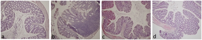
Histological evaluation of colon inflammatory cell infiltration in female and male BTBR mice
(a) Colon tissue from mC57 mice showing absence of inflammatory cells.
(b) Colon tissue of mBTBR group showing leukocyte infiltration in the mucosa and submucosa. (part of intestines)
(c) Colon tissue of fC57 group, showing absence of inflammatory cells.
(d) Colon tissue from fBTBR group showing moderate leukocyte infiltration in the mucosa.(part of intestines)
Histological evaluation of colon inflammatory cell infiltration in female and male BTBR mice. [Digital image]. (2017, March 28). Retrieved April 23, 2017, from https://www.ncbi.nlm.nih.gov/core/lw/2.0/html/tileshop_pmc/tileshop_pmc_inline.html?title=Click%20on%20image%20to%20zoom&p=PMC3&id=5368984_srep45356-f4.jpg
